Alkali Burn-Induced Keratitis Model

Alkali Burn-Induced Keratitis Model
Huateng Bio provides validated alkali burn-induced keratitis models in rabbits. Features 21-day neovascularization tracking, slit-lamp scoring, and ISO-compliant histopathology. Ideal for ocular drug development. Download protocols.
Model Description
Corneal alkali burns induce progressive ocular damage through dual physicochemical mechanisms:
1. Immediate tissue necrosis: NaOH-mediated epithelial dissolution and stromal collagen degradation
2. Chronic pathology: Inflammatory cell infiltration, neovascularization, and corneal ulceration
Our model replicates key clinical features of severe keratitis:
- Corneal opacity (≥Grade 3 on Fantes scale)
- Neovascular invasion (>5 vessels/quadrant at Day 21)
- Epithelial defect persistence (fluorescein staining ≥4mm²)
Clinical Relevance:
✓ Mimics human chemical burns (workplace/domestic accidents)
✓ Supports therapeutic testing: Anti-angiogenics, anti-inflammatories, and corneal repair agents
Applications
• Anti-inflammatory drug efficacy evaluation (corticosteroids, NSAIDs)
• Corneal neovascularization inhibition studies
• Stem cell therapy validation for epithelial regeneration
• Collagen cross-linking protocol optimization
Modeling Protocol —— NaOH-Induced Corneal Burn
1. Pre-op Preparation:
- Anesthetize rabbits (ketamine 35 mg/kg + xylazine 5 mg/kg)
- Confirm corneal integrity via slit-lamp exam
2. Burn Induction:
- Apply 1% NaOH-saturated filter paper (5mm diameter) to central cornea for 30 seconds
- Rinse with 200mL saline (pH 7.4)
3. Post-op Care:
- Levofloxacin eye drops TID ×3 weeks
- Weekly clinical scoring: Opacity/neovascularization/fluorescein retention
Validation & Testing
|
Category |
Parameters |
|
Slit-Lamp Examination |
• Opacity grading (0-4 scale) |
|
Fluorescein Staining |
Defect area quantification (ImageJ analysis) |
|
Histopathology |
H&E staining: Epithelial thickness ∙ Inflammatory cell count |
|
Molecular Analysis |
ELISA: VEGF, IL-1β, TNF-α ∙ MMP-9 zymography |
Data
(1) Corneal opacity: The corneal opacity score is the sum of the burn spot score and the opacity score outside the burn spot. Under a slit lamp microscope, observe and score the corneal opacity of the animals except those in the negative control group.
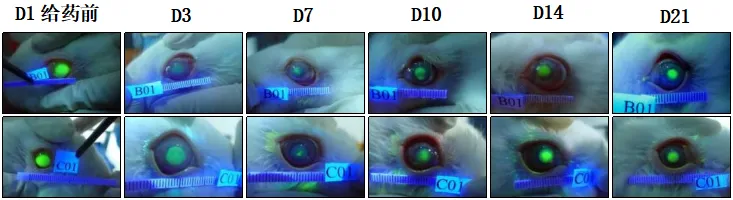
Fluorescein sodium staining image of corneal alkaline burn
(2) Histopathological Analysis
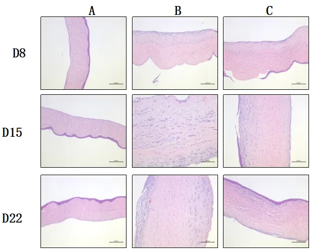
Histopathological images of rabbit corneal tissue at 8, 15, and 22 days after corneal alkaline burn modeling
Related Animal Models
Explore other gene-edited animal models that complement our porcine research platforms