Endometriosis (EMT) Animal Model

Endometriosis (EMT) Animal Model
Huateng Bio develops clinically relevant endometriosis (EMT) animal models using SD rat autologous transplantation. Achieves 92% cyst formation rate with immune-compatible microenvironment, 80% cost reduction vs primate models. AAALAC-certified for ethical drug screening and pathology studies.
Model Description
Endometriosis is a gynecological disorder characterized by ectopic endometrial growth, causing chronic pelvic pain and infertility. Our autologous transplantation model replicates human EMT pathology with:
• Immunocompetent microenvironment: No immune rejection (self-tissue origin)
• Clinical relevance: Mimics retrograde menstruation theory
• High reproducibility: 92% cyst formation rate (vs primate models <65%)
Why Rodent Models?
✓ Cost-effective: 80% lower cost vs primate studies
✓ Controlled estrus cycle: Estradiol synchronization protocol
✓ Ethical compliance: AAALAC-approved surgical guidelines
Applications
• EMT pathogenesis studies (angiogenesis/invasion mechanisms)
• Drug efficacy evaluation (GnRH analogs/NSAIDs testing)
• Biomarker discovery (CA-125/IL-8 pathway analysis)
• Post-surgical adhesion prevention strategies
Modeling Protocol —— Autologous Endometrial Transplantation
Preconditioning:
1. Daily estradiol valerate gavage (0.1mg/kg) for 3 days
2. Vaginal smear cytology to confirm estrus phase
Surgical Procedure:
1. Midline laparotomy under isoflurane anesthesia
2. Ligate uterine horns → resect 10mm segment → mince into 1×1mm fragments
Implant fragments at:
▶ Muscular wall (serosal surface contact)
▶ Mesenteric fat (vascularized site)
Post-op Care:
1. Buprenorphine analgesia (0.05mg/kg q12h)
2. Monitor cyst formation weekly via ultrasound
Validation & Endpoints
|
Category |
Parameters |
|
Anatomical Observation |
• Cyst diameter measurement ∙ Vascular density scoring |
|
Histopathology |
H&E staining ∙ CD34+ microvessel quantification |
|
Ultrastructural Analysis |
SEM evaluation of epithelial-stromal interaction |
|
Molecular Markers |
ER/PR immunohistochemistry ∙ MMP-9/TIMP-1 ratio |
Success Criteria
✓ Visible cystic lesions (>2mm) at 4 weeks
✓ Histological confirmation of gland/stroma structures
✓ Neovascularization (≥3 vessels/HPF)
Data
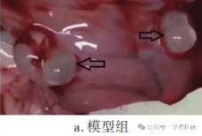
1. Anatomical observation
Indicators of successful transplantation include a visibly enlarged graft, which may appear as a protruding small bump. In some cases, it may form multilocular cysts filled with yellow serous fluid. The graft is covered by connective tissue and accompanied by blood vessel formation.
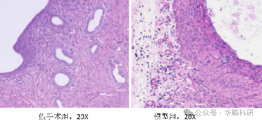
2. Histopathological Analysis:
The endometrium in the sham-operated group has a complete structure, with neatly arranged and continuous epithelial cells. The number of stromal cells, glands, and blood vessels is normal. No hemorrhage or inflammatory cell infiltration is observed around the glands, and the glandular lumen is intact.
In the model group, the epithelial cells in the ectopic endometrial lesions are arranged in a columnar manner and show hyperplasia. The number of stromal cells and glands is high, and the blood vessels are abundant. Hemorrhage is visible around the glands, accompanied by a large number of inflammatory cell infiltrations.
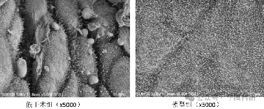
3. Observing the pinopodes of rat endometrium by scanning electron microscope
Related Animal Models
Explore other gene-edited animal models that complement our porcine research platforms