Rats Cranial Defect Model
Rats Cranial Defect Model
The cranial defect model is a widely used experimental tool for studying bone regeneration mechanisms, evaluating biomaterials, and assessing the efficacy of osteogenic agents.
Model Description
The cranial defect model is a widely used experimental tool for studying bone regeneration mechanisms, evaluating biomaterials, and assessing the efficacy of osteogenic agents. This model involves creating standardized skull defects through surgical drilling, making it suitable for investigating intramembranous ossification, testing the osteoinductive properties of biomaterials, and screening drugs for bone regeneration applications. It provides essential experimental data for research in tissue engineering and regenerative medicine.
Applications
- Mechanisms of bone regeneration
- Evaluation of biomaterials
- Screening of bone regenerative drugs
Modeling Methods
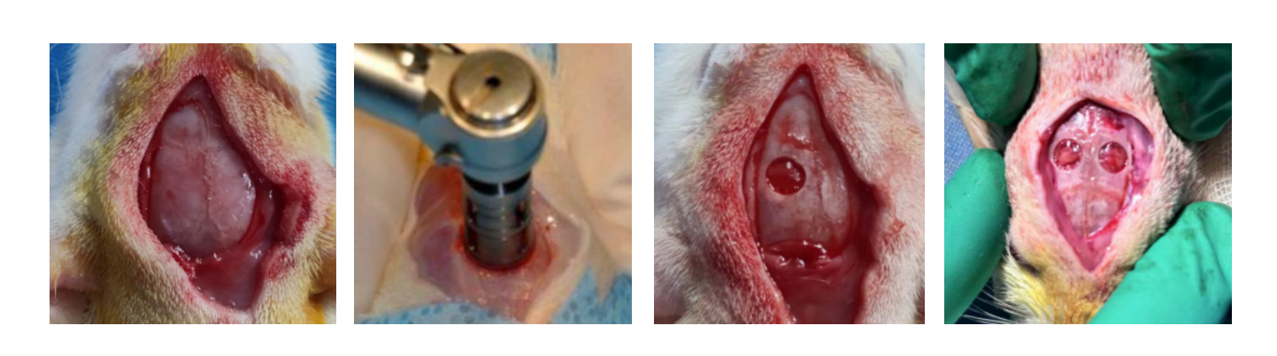
Anesthetized SD rats are prepared by shaving the cranial region, followed by skin disinfection and sterile draping. The animals are fixed in the prone position, and a midline skin incision is made to expose the periosteum. The muscle layer is carefully dissected to fully expose the cranial surgical site. Using a low-speed drill with a 5mm diameter trephine bur, full-thickness cranial defects are created bilaterally along the sagittal suture. Care is taken to avoid damaging the dura mater during the procedure.
After achieving complete hemostasis, experimental materials are implanted into the defect sites, ensuring a 2-3mm margin of coverage around the defects. The incision is then closed in layers, completing the surgical procedure.
Testing Items
- Micro-CT analysis of new bone tissue: bone volume fraction (BV/TV), trabecular number (Tb.N), trabecular thickness (Tb.Th), and trabecular spacing (Tb.Sp)
- Histopathological analysis of the cranial defect area
Related Animal Models
Explore other gene-edited animal models that complement our porcine research platforms