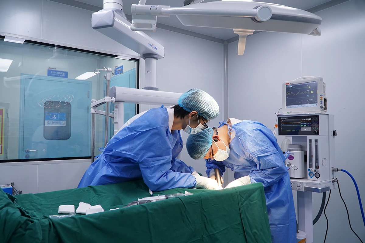
animal models for respiratory disease research
With deep capabilities in respiratory function measurement, bronchoscopy, advanced imaging, and molecular analysis, we provide translationally relevant models to support your drug discovery and mechanistic studies in respiratory medicine.
|
Chronic Obstructive Pulmonary Disease (COPD) Model |
An integrative model induced by chronic cigarette smoke exposure, often combined with elastase instillation to emulate both emphysema and chronic bronchitis. |
|
Chronic Bronchitis Model |
Induced by repeated exposure to irritants like sulfur dioxide (SO₂) or cigarette smoke, characterized by mucus hypersecretion and airway inflammation. |
|
Pulmonary Emphysema Model |
Primarily induced by intratracheal elastase administration or genetic modifications, leading to progressive alveolar destruction and airspace enlargement. |
|
Airway Remodeling Model |
Established via chronic allergen challenge or TGF-β overexpression to study structural changes like fibrosis and smooth muscle hypertrophy. |
|
Acute Lung Injury (ALI) Model |
Commonly induced by lipopolysaccharide (LPS) administration or ventilator-induced injury to study diffuse alveolar damage. |
|
Acute Respiratory Distress Syndrome (ARDS) Model |
A more severe form of ALI, often modeled using a combination of insults like LPS and ventilator-induced injury. |
|
Pulmonary Edema Model |
Induced by oleic acid intravenous injection or high-pressure ventilation to investigate mechanisms of vascular leakage and fluid accumulation. |
|
Pulmonary Ischemia-Reperfusion Injury Model |
Created by temporarily clamping the pulmonary artery (e.g., in transplant settings), crucial for studying post-surgical lung damage. |
|
Pulmonary Hypertension (PH) Model |
The classic model induced by a single injection of monocrotaline (MCT) in rats, causing vascular remodeling and increased pressure. |
|
Hypoxia-Induced Pulmonary Hypertension Model |
Induced by housing animals in a sustained hypoxic chamber, ideal for studying the role of hypoxia in PH development. |
|
Pulmonary Embolism Model |
Created by intravenous injection of autologous blood clots or microspheres to physically obstruct pulmonary arteries. |
|
Pneumonia Model |
Established by direct airway inoculation with pathogens like Pseudomonas aeruginosa or Streptococcus pneumoniae. |
|
Pulmonary Tuberculosis Model |
Utilizes infection with Mycobacterium tuberculosis in susceptible mouse strains or guinea pigs to test vaccine and drug efficacy. |
|
Pneumonia Model (Viral) |
Can be established using relevant viruses (e.g., influenza, SARS-CoV-2) to model viral pathogenesis and immune responses. |
|
Asthma Model |
The standard model involves sensitization and airway challenge with allergens like ovalbumin (OVA) or house dust mite (HDM). |
|
Airway Allergic Disease Model |
Similar to the asthma model, focusing on the allergic sensitization phase and Th2-driven inflammation. |
|
Rhinitis Model |
Induced by local allergen challenge in the nasal mucosa, used for studying allergic rhinitis and nasal inflammation. |
|
Pulmonary Fibrosis Model |
The gold-standard model induced by a single intratracheal instillation of bleomycin in mice, leading to progressive scarring. |
|
Acute Lung Injury (ALI) Model |
Can also be used to study the early inflammatory phase that may precede fibrosis. |
|
Lung Cancer Model |
- Chemically-induced: Urethane injection for spontaneous lung tumor development. |
|
Lung Transplantation Model |
A microsurgical model in rodents for studying allograft rejection, immunosuppression, and ischemia-reperfusion injury. |
|
Hypoxia Exposure Model |
Animals are housed in hypoxic chambers to study physiological adaptations or pathologies related to high-altitude or low-oxygen conditions. |
|
Tracheal/Airway Stenosis Model |
Created by mechanical or chemical injury to the trachea, used for researching wound healing and restenosis prevention. |
|
Sinusitis Model |
Induced by bacterial inoculation or sinus ostium obstruction to model acute or chronic sinusitis. |
Global Compliance: AAALAC-accredited facilities with IACUC protocols aligned with EU Directive 2010/63/EU and USDA standards.
Advanced Imaging: DSA, IVUS, angiography, and micro-CT for real-time device performance tracking.
Custom Model Development: Species-specific animal models for complex disease states.
Histopathology Suite: SEM/TEM analysis with GLP-grade reporting for PMDA/CE Mark submissions.