Femoral Condyle Defect Model
Femoral Condyle Defect Model
Huateng Bio offers rabbit femoral condyle defect models for bone regeneration research. Features 5mm critical-sized defects, Micro-CT quantification, and human-like remodeling. Ideal for orthopedic implant validation.
Model Description
The rabbit femoral condyle defect model is a gold-standard preclinical tool for evaluating bone regeneration strategies and orthopedic implant performance. Leveraging rabbits' human-like skeletal remodeling capacity and large bone dimensions, this model enables:
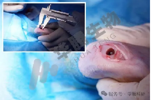
- Critical-sized defect standardization: 5mm diameter × 10mm depth cylindrical defects
- Implant compatibility testing: Accommodates human-scale grafts (hydroxyapatite, PCL scaffolds)
- High reproducibility: 95% defect consistency via guided osteotomy
Key Advantages:
✓ Anatomical relevance: Trabecular bone density comparable to human femoral condyles
✓ Controlled healing phases: 6-12 week monitoring window
✓ Multi-modal validation: Micro-CT, biomechanics, and histopathology integration
Applications
• Bone graft material efficacy testing (ceramics, polymers, composites)
• 3D-printed scaffold osseointegration studies
• Growth factor delivery system evaluation (BMP-2, VEGF)
• Non-union defect mechanism research
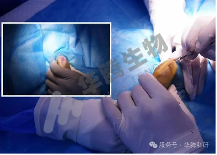
Modeling Protocol —— Surgical Defect Creation
1. Preoperative Prep:
- Anesthetize rabbits (ketamine/xylazine 35/5 mg/kg)
- Shave and disinfect the lateral femoral condyle region
2. Defect Generation:
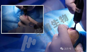
- Lateral parapatellar incision to expose the femoral condyle
- Create a 5mm diameter × 10mm depth cylindrical defect using a surgical osteotomy drill(500 RPM, saline irrigation)
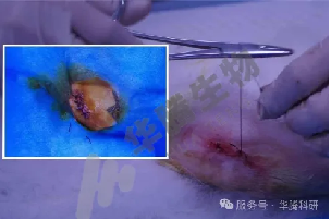
3. Closure:
- Layer-by-layer suture (4-0 Vicryl)
- Post-op analgesia (meloxicam 0.2 mg/kg)
Data
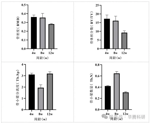
1. Micro CT :
Bone Mineral Density (BMD), Bone Volume Fraction (BV/TV), Trabecular Separation (Tb.Sp), Trabecular Number (Tb.N).
Related Animal Models
Explore other gene-edited animal models that complement our porcine research platforms